- 01: Introduction
- 02: History
- 03: Propellants, Firearms, and Ammunition Development
- 04: Modern Firearms Manufacture
- 05: Small Arms Ammunition
- 06: Evidence Handling Procedures
- 07: Equipment and Instrumentation
- Introduction
- Objectives
- AFTE Knowledge and Ability Factors
- Measurement, Standards, and Accuracy
- Weight, Force, and Their Measurement
- Dimensional Data and Measurement Tools
- Muzzle Velocity of Bullets
- Stereo and Comparison Microscopes
- Small Tools and Supplies
- Imaging Equipment
- Field Support Equipment
- Computer-Based Technologies
- Firing Facilities
- Selected Bibliography
- 08: Examination of Firearms
- 09: Cartridge and Shotshell Examination
- 10: Characterization and Evaluation of Fired Projectiles
- 11: Bullet Comparison and Identification
- 12: Gunshot Residue and Distance Determination
- 13: Toolmark Identification
- 14: Communicating Results
- Resources


Compound Microscopy
Home > Equipment and Instrumentation > Stereo and Comparison Microscopes > Compound Microscopy
)
Basic diagram of a compound
light microscope
Credit for the invention of the compound microscope is often given to Zacharias Janssen, a spectacle maker from Middleburg, Holland (1590s). In 1610, Galileo announced his version, followed by many other prominent pioneers in the field of optics. They found that when two simple convex lenses with specific characteristics were placed at the ends of a tube with a defined distance between them, much greater magnifications could be achieved. This constituted the first compound microscope – one containing more than a single lens. The lower lens (nearest to the object being observed) was called the objective lens; the lens closest to the observer was called the eyepiece or ocular lens. When the entire assembly was raised or lowered, the object being observed could be brought into focus. When the object is brought into clear focus, an inverted image is formed by the objective lens at a point within the principle focus of the ocular lens. In turn, this image becomes the object for the ocular lens. This results in an even larger image for the observer. These early compound microscopes typically had magnifications of 3x to 9x.
This was a great leap forward, but there were optical problems with both single lens and compound microscopy.
The problems included
- dimensional distortion,
- color inaccuracies (especially at the edges of the round convex lenses used in the systems),
- inverted images.
These and other problems were corrected by the use of additional combinations of complex concave and convex lenses added to the microscope lens tube. By 1873, German Ernst Leitz had invented the revolving nosepiece for quickly rotating objective lenses.
Stereomicroscopy
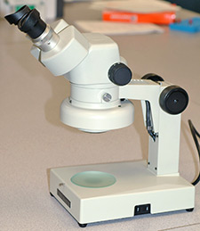
Stereomicroscope
Stereomicroscopy is based on the use of physically joined compound microscopes (one for each eye) in the examination of single objects. Sometimes a single objective lens is shared by both eyepieces. This approach yields a three-dimensional (stereo) view of an object, such as a fired bullet being evaluated prior to microscopic comparison with another bullet. It is particularly useful when combined with an optical zoom feature, as is common in many stereomicroscopes. The stereomicroscope is useful during any phase of forensic firearms work, including the next level of visible light microscopy – comparison microscopy.